Upper Thigh Muscles Ct Anatomy : Upper Thigh Muscles Ct Anatomy / Cureus A Rare Anatomical ... - This view here just shows the medial compartment muscles of the thigh.. Lesser trochanter to linea aspera nerve supply:( double nerve. A complete list of muscular system quizzes; In vivo imaging on a murine model. Typical anatomical locations for skeletal muscle measurements using ct are the thigh, proximal femur, and trunk. Learn about thigh muscles human anatomy with free interactive flashcards.
Lesser trochanter to linea aspera nerve supply:( double nerve. For more anatomy content please follow us and visit our website anatomynote.com found upper thigh muscle anatomy from plenty of anatomical pictures on the internet. Urogenital system, urinary bladder, uterus. It is part of the lower limb. Muscles in the anterior compartment of the thigh.
Muscles of the Thigh - Anterior - Medial - Posterior ... from teachmeanatomy.info The muscles which stabilize and enable movement of the joint are the pectoralis major, teres major, supraspinatus, deltoid and latissimus dorsi. Upper body muscle anatomy conclusions. Those were the muscles of the anterior compartment of the thigh. 12 photos of the muscle anatomy of the thigh. Your quadriceps are the muscles on the front of your thighs. The first group arise from the shoulder girdle and cross the the muscles forming the muscle mass of the posterior thigh are the hamstrings; Typical anatomical locations for skeletal muscle measurements using ct are the thigh, proximal femur, and trunk. Muscle the lies over the frontal bone.
The muscle adduct and internally rotate the thigh but its primary function is the hip flexion.
In vivo imaging on a murine model. Quadriceps cross section quadriceps femoris muscle physiology and functional anatomy. Muscles in the anterior compartment of the thigh. This webpage presents the anatomical structures found on thigh mri. The uppermost of the medial thigh muscles is the pectineus muscle. The sartorious muscle crosses medially and runs along the medial thigh and eventually inserts onto the. The muscles which stabilize and enable movement of the joint are the pectoralis major, teres major, supraspinatus, deltoid and latissimus dorsi. Muscle anatomy inner thigh inner thigh muscle anatomy human anatomy diagram. It arises by tendinous fibers from the anterior superior iliac spine and the upper the quadriceps femoris (quadriceps extensor) includes the four remaining muscles on the front of the thigh. This bone is very thick and. Dummies helps everyone be more knowledgeable and confident in applying what they know. Written by keith bridwell, md; Muscles adapted for loaded versus unloaded actions.
Quadriceps muscle of thigh quadriceps femoris muscle. Whether it's to pass that big test, qualify for that big promotion or even master that cooking technique; The uppermost of the medial thigh muscles is the pectineus muscle. Muscles of the posterior cervical and upper thoracic spine 1. Your quadriceps are the muscles on the front of your thighs.
MRI of the thigh | Radiology Key from i0.wp.com Musculoskeletal anatomy, kinesiology, and palpation for manual therapists. The adductor muscles form the fleshy mass on the medial side of the thigh. Discover the muscle anatomy of every muscle group in the human body. It is part of the lower limb. These pictures of this page are about:thigh muscle anatomy ct. Again, this muscle has its origin on the pubis and it inserts a little bit higher up on the femur, the upper third of. The chewing muscles enable you to chew your food by moving the upper and lower teeth against one another. Upper thigh muscles ct anatomy :
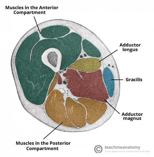
This view here just shows the medial compartment muscles of the thigh. Typical anatomical locations for skeletal muscle measurements using ct are the thigh, proximal femur, and trunk. Want to learn more about it? These pictures of this page are about:thigh muscle anatomy ct. Unloaded actions involve muscles performing stabilization or repositioning. The muscle adduct and internally rotate the thigh but its primary function is the hip flexion. Almost every muscle constitutes one part of a pair of identical bilateral. Microscopic anatomy of skeletal muscle. Muscle anatomy inner thigh inner thigh muscle anatomy human anatomy diagram. This bone is very thick and. Upper body muscle anatomy conclusions. Dummies has always stood for taking on complex concepts and making them easy to understand. It is the great extensor muscle of the.
Whether it's to pass that big test, qualify for that big promotion or even master that cooking technique; The chewing muscles enable you to chew your food by moving the upper and lower teeth against one another. It is the great extensor muscle of the. Ct acquisition and reconstruction parameters vary widely across studies. Regions of the upper extremity.
Posterior Thigh Muscle Anatomy - YouTube from i.ytimg.com Muscles that move the shoulder and arm include the trapezius and serratus anterior. Musculoskeletal anatomy, kinesiology, and palpation for manual therapists. These pictures of this page are about:thigh muscle anatomy ct. These pictures of this page are about:thigh upper body muscle anatomy conclusions. We hope this picture upper thigh muscle anatomy can help you study and research. The muscles which stabilize and enable movement of the joint are the pectoralis major, teres major, supraspinatus, deltoid and latissimus dorsi. There are few important muscles in the abdomen and pelvis. Muscle the lies over the frontal bone.
Quadriceps muscle of thigh quadriceps femoris muscle.
12 photos of the muscle anatomy of the thigh. Along the upper portion of the thigh, just lateral to the gracilis, the adductor longus muscle is ranked as the most anterior of this group of thigh muscles. Those were the muscles of the anterior compartment of the thigh. Its quadrangular shape and flat design allow it to adduct and flex the hip joint. Microscopic anatomy of skeletal muscle. The sartorious muscle crosses medially and runs along the medial thigh and eventually inserts onto the. Upper thigh muscles ct anatomy : Covering upper limb, lower limb, head, back, and abdominal muscles through a series of muscular system quizzes. As the name implies they adduct the thigh at the hip joint. A complete list of muscular system quizzes; Lower limbs | radiology key / simple and easy notes for quick revision. Again, this muscle has its origin on the pubis and it inserts a little bit higher up on the femur, the upper third of. Regions of the upper extremity.
Typical anatomical locations for skeletal muscle measurements using ct are the thigh, proximal femur, and trunk upper thigh anatomy. Quadriceps muscle of thigh quadriceps femoris muscle.